A network science cartography of cognitive control system dynamics

For more details on these Figures, see the full report here.

Understanding the neural mechanisms of cognitive control is essential to understanding the relation of human brain function and behavior. Cognitive control refers to processes supporting goal-directed behavior and cognition. A rich collection of executive functions are under the purview of cognitive control, including (but not limited to): decision making, problem solving, endogenous attention, mood regulation, motivation, working memory, and many forms of learning. In fact, many modern neuroscientists consider the term cognitive control to be synonymous with executive functioning. Importantly, cognitive control is also thought to be negatively impacted in many mental illnesses, which altogether makes it an imperative topic for a varied cross-section of researchers. 

Prior research has identified large-scale neural systems collectively termed cognitive control networks. Of these, two prominent control networks include the frontoparietal network (FPN) and the cingulo-opercular network (CON). Brain regions of cognitive control networks are thought to enact control by large-scale network interactions that are flexible to the current goal or task context (Waskom et al., 2014). This framework is built upon the biased competition theory (Desimone et al., 1990, 1995), then later the guided activation theory (Miller and Cohen, 2001), and then more recently, the flexible hub theory (Cole et al., 2013). The flexible hub theory was developed by our lab and posits that well-connected hub regions exhibiting global connectivity changes (across tasks) are critical to adaptive task control.

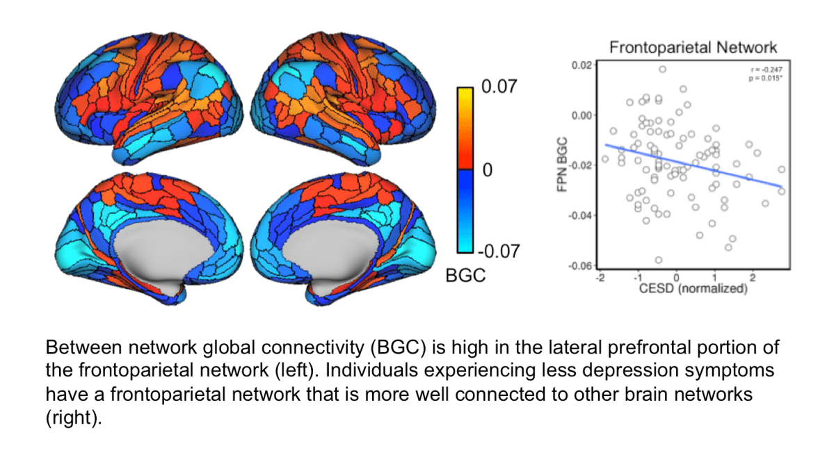
Prior work has delineated the cognitive control related properties of FPN regions well. However, control properties of CON regions are less established. In our recent paper published in The Journal of Neuroscience, we examined the neural mechanisms of both the FPN and CON by quantifying each of their large-scale network interactions during a high-control-demand cognitive paradigm (as in Cole et al., 2010, 2013; Ito et al., 2017). We used fMRI data acquired during both resting and task states for 100 healthy adults. Estimates of functional connectivity were used to probe network interactions. This was based on the idea that these estimates capture interregional information flow in a functionally-relevant manner. To characterize the dynamic, functional properties of control networks, we applied network science tools (including a novel metric termed “network partition deviation”), decoding analyses, and developed a multidimensional functional cartography (or “mapping”; panel C in above Figure). 

Expanding upon the flexible hub theory (and related theories mentioned above), we found FPN regions exhibited a “flexible coordinator” mechanism (panel A in above Figure). The “flexible” aspect of this characterization was observed in between-network connectivity patterns demonstrating high variability across task states. This suggests that FPN regions exhibit network interactions that are continuously adaptive to task goals. The “coordinator” aspect of this characterization was observed in FPN’s within-network configuration being maintained across task states. This internal stability could facilitate the binding of stimulus information with task-rule information during cognitive computations.

We found that the CON exhibited mechanisms of cognitive control that were complementary to the FPN: namely, acting as a “flexible switcher” (panel B in above Figure). Here, the “flexible” portion was demonstrated by CON regions exhibiting transient, task-relevant connectivity changes. These connectivity changes were observed as disbandments from their resting-state configuration, constituting the “switcher” portion of the CON’s characterization. This suggests that CON is capable of augmenting the processing of goal-relevant regions (or networks) by lending them processing resources. This proposed role for CON in controlled processing expands upon findings that suggest it specifies overall task-set modes (Dosenbach et al., 2007; Power and Petersen, 2013; Sadaghiani and D’Esposito, 2015).

Taken together, this account highlights the FPN and CON enacting cognitive control in a complementary manner. But what may be the benefit of the control networks exhibiting these specific properties? An idea central to the theories mentioned above is competition. Neural representations compete for resources: in any given moment (i.e., during a task), representations of stimuli, actions, and/or thoughts are all vying for the limited processing resources of the brain. It is thought that top-down signals from control network regions can shift this competition in favor of neural representations that are relevant to the task or goal at hand. This is the essence of cognitive control. To this end, FPN connectivity patterns appear important for flexibly coordinating top-down biases across task states. However, interference (from goal-irrelevant representations) is still possible. The flexible switching of CON connections can step in to lend resources to the goal-relevant systems and help them win competitions. Thus, the FPN and CON may be acting in-concert toward effective controlled processing. Moreover, this account allows for the possibility that the control networks are managing a tradeoff in computational demand.

Looking forward, we hope that this work is built upon by incorporating other task paradigms, clinical cohorts, and other applications targeting cognitive control. Methodological considerations can be taken into account, such as improving the estimation and interpretation of functional connectivity. The cartographic approach could be applied to other functions of interest (and accordingly, task paradigms with other outcomes), such as visual processing or attention. Moreover, recent findings across a variety of research questions point to a broad distinction between sensorimotor versus cognitive network mechanisms (e.g., Bassett et al., 2013). Our pipeline could be implemented as a tool for exploring these distinctions further. 

Check out the full report here.

Less depression for those with a better-connected frontoparietal brain network

Does the fronto-parietal cognitive control network (FPN) function as an “immune system of the mind?” This hypothesis was developed based on work in our lab (Cole, Repovš, & Anticevic, 2014) and others, suggesting that the FPN consists of flexible hubs.

Flexible hubs are regions in the brain that are especially well connected to other systems in the brain that can rapidly modify those functional connections based on current goals.

Based on this information and evidence suggesting that FPN connections are altered in a number of mental disorders we hypothesized that the FPN could serve to regulate dysfunctional communication between other brain systems that may occur in mental disorders. In a sense, the FPN could be correcting problems with brain communication that occur in mental disorders. This would help bring the brain back to a more healthy state and decrease mental health symptoms.

One of the important aspects of the flexible hub nature of the FPN is that it is well connected to other areas of the brain. This high degree of connectivity would allow the FPN to be able to influence a large number of other brain networks and adjust problems similar to how a thermostat adjusts the temperature of your home.

We recently published an article in Network Neuroscience that tested the idea that the flexible hub properties of the FPN may serve a protective role against mental health symptoms.

All people vary in their level of mental health. Even people who have not been diagnosed with a disorder can show different degrees of symptoms. Exploring these differences may help us better understand mental illness and problems that don’t reach the criteria for a disorder but may still negatively impact peoples’ quality of life. We predicted that individuals with greater connectivity between the FPN and other brain networks would report fewer mental health symptoms. We tested this hypothesis by assessing brain connectivity with resting-state fMRI in 96 young adults who had not been diagnosed with a mental health disorder. We also had participants complete a questionnaire that assessed how frequently they experienced different symptoms related to depression.

Briefly, we found that lateral prefrontal portions of the FPN are well connected to other brain systems. We also found that individuals with a better-connected FPN experienced depression symptoms less frequently. These findings suggest that the organization of global brain networks is important for maintaining mental health, even in individuals who have not been diagnosed with a mental disorder. This supports the hypothesis that the FPN may play a role in regulating mental health symptoms as they arise through goal-directed feedback.

You can find the full report at Network Neuroscience, here.

A network mechanism for the transfer of cognitive information between brain regions

blog_fig

Historically, cognitive neuroscience has mapped cognitive processes onto specific brain areas. However, it has more recently become evident that the brain processes information in a distributed manner, with networks of brain regions working together to process cognitive information. We recently published an article in Nature Communications addressing this issue, investigating how cognitive information in one brain region is transferred to another brain region through routes described by intrinsic network architecture.

Briefly, our results suggest that the human brain’s intrinsic network architecture (i.e., the network architecture extracted while a person is resting in a functional magnetic resonance imaging scanner) forms the foundation upon which distributed cognitive processing occurs. Using the brain’s intrinsic network architecture, we could predict how patterns of activity related to active cognitive processing are propagated between brain regions and networks. We first validated this principle using a computational model, where we simulated a resting brain and showed how patterns of intrinsic connectivity can predict how information is transferred during simulated cognitive tasks. We then collected empirical fMRI data while human subjects performed an array of cognitive tasks, showing that we could predict cognitive information in distributed brain areas using their intrinsic (resting) network architecture. In follow-up analyses, we found that cognitive control networks (brain networks with hub-like properties) played a disproportionate role in transferring information.

The resting brain has already been shown to be able to predict how brain regions will activate during tasks (Tavor et al., 2016; Cole et al., 2016). We built on these findings to show that patterns of connectivity between brain regions describe the channels of activity flow between those regions that carry cognitive information. This allowed us to infer that information in distributed parts of the brain are related to each other through an underlying, intrinsic network architecture. We hope these findings will spur new investigations into computational roles that intrinsic brain networks play in information communication and transformation.

Here’s a link to the article: http://rdcu.be/wQ1M

Citations:

Ito, T., Kulkarni, K. R., Schultz, D. H., Mill, R. D., Chen, R. H., Solomyak, L. I., & Cole, M. W. (2017). Cognitive task information is transferred between brain regions via resting-state network topology. Nature Communications. https://doi.org/10.1038/s41467-017-01000-w

Tavor, I., Jones, O. P., Mars, R. B., Smith, S. M., Behrens, T. E., & Jbabdi, S. (2016). Task-free MRI predicts individual differences in brain activity during task performance. Science, 352(6282), 1773–1776. https://doi.org/10.1126/science.aad8127

Cole, M. W., Ito, T., Bassett, D. S., & Schultz, D. H. (2016). Activity flow over resting-state networks shapes cognitive task activations. Nature Neuroscience. https://doi.org/10.1038/nn.4406


Frequently asked questions about activity flow mapping, Part 2

coleetal2016fig1a
The activity flow mapping procedure. See Cole et al. (2016; Nature Neuroscience) for more info.

I recently posted a brief summary of the Cole et al. (2016; Nature Neuroscience) paper, along with answers to some frequently asked questions. I’m including answers to additional frequently asked questions here. This gets into more of the lesser-known implications of the activity flow mapping findings.

What can explain the success of activity flow mapping at a fine-grained mechanistic level? What could possibly explain the ability to predict held-out activations at a distance?

The most parsimonious explanation is that fMRI BOLD signals strongly reflect action potential propagation. Action potential propagation is the only known mechanism for the brain to synchronize distal locations (i.e., implement activity flow). Note that this interpretation is still consistent with the previous suggestion that fMRI BOLD primarily reflects local field potentials, given that local field potentials are caused by incoming action potentials. This direct causal link between local field potentials and action potentials suggests it is likely quite rare for local field potential variance to be dissociated from incoming action potential variance at the neural level. Even if sub-threshold local field potential variance drives fMRI signals frequently, however, functional connectivity estimates likely isolate the variance that is most strongly tied to action potentials. This is because, as mentioned, action potentials are the only known (non-artifactual) mechanism for creating the long-distance correlations observed with fMRI functional connectivity. I think this is an important point to consider moving forward when interpreting fMRI functional connectivity estimates.

Given the dependence of functional connectivity estimates on action potentials, the success of the activity flow mapping approach suggests that task-evoked activations likely also largely reflect action potentials. However, it is possible that an activation in a given region is largely reflecting local field potentials (i.e., there is little spiking output from that region), with those local field potentials being driven by action potentials from other regions.

Given that you are using fMRI (which relates to neural-activity-induced changes in blood flow), can you rule out the possibility that the “flow” in activity flow mapping is actually blood flow?

Vasculature-based blood flow likely influences highly local correlations in fMRI BOLD signals. We had actually considered this issue in our methodology, under the assumption that such blood flow effects would likely not extend beyond 9 millimeters. Thus, activity flow estimates less than 9 millimeters were excluded. Note that this is likely overly cautious, since blood flow effects are unlikely to extend as far as 9 millimeters (it’s effects are likely restricted to within ~2.5 millimeters, with the largest effects due to draining veins). Thus, the results suggest the most likely (non-artifactual) cause of fMRI BOLD correlation over distances greater than ~2.5 millimeters is activity flow via action potential propagation.

You talk of rest and task as if they’re different things, yet a premise of the paper is that it’s the same networks that drive both.  Why is rest not a task?

I definitely think of rest as a sort of (less constrained) cognitive task. We focused on resting-state fMRI data here for several pragmatic reasons. First, this is the convention in the literature, largely because it’s easier to collect resting-state data than task data. Second, PET imaging has been used to show that rest is the state with the lowest overall metabolic demanding conscious state. This suggests it may be the best-available cognitive baseline. That said, we are really using resting-state functional connectivity as a proxy to true intrinsic functional connectivity, which would not be influenced by the idiosyncrasies of resting state. This might involve deriving a cross-state functional connectivity map, as we did in a recent study, such that the resulting map was not strongly biased by any single mental state.

How is it possible for activity flow mapping to accurately predict task activations when we know resting-state functional connectivity estimates are corrupted via a large number of artifacts, especially motion-related artifacts?

The activity flow mapping results should be surprising if you believe fMRI functional connectivity is corrupted by artifacts more than fMRI task activations (which some studies have suggested). Task fMRI activations are thought to be largely immune to artifacts such as motion that influence functional connectivity estimates, due to the benefit of experimental control dissociating task-related activations from the timing of various artifacts. The success of the activity flow mapping approach demonstrates that these artifacts may not be as bad as we thought, especially as far as correspondence to task activation is concerned. Note, however, that activity flow mapping worked better when multiple regression was used to estimate connectivity – a method thought to reduce the influence of such artifacts on the data. Nonetheless, the approach worked quite well using the standard Pearson correlation approach to functional connectivity, suggesting much of the variance included in standard resting-state functional connectivity is likely non-artifactual.


Frequently asked questions about activity flow mapping (Cole et al., 2016; Nature Neuroscience)

coleetal2016fig5
Figure 5 from Cole et al. (2016; Nature Neuroscience)

We recently published a paper in Nature Neuroscience that we’re quite excited about. It’s titled “Activity flow over resting-state networks shapes cognitive task activations“. Here we are going to go over some lesser-known aspects of the study. We will begin, however, with a basic overview of the results.

Briefly, we sought to gain some clarity on what the cognitive relevance of resting-state networks might be. These networks have been studied extensively with hundreds of papers now being published each year using resting-state fMRI. But what do they have to do with cognition? In particular, how do resting-state networks mechanistically relate to cognitive functions?

We hypothesized that activity flow – the movement of activation amplitudes between brain regions – is the network mechanism that explains the relevance of resting-state networks to cognitive activations (and therefore to cognition). We found strong support for this hypothesis using both a simple computational model and empirical fMRI data.

I’ll now switch to a frequently asked questions (FAQ) format, digging into some lesser-known conclusions from the study:

Wasn’t this already known based on previous studies showing that resting-state networks are similar to a variety of task-evoked activation patterns?

Those studies certainly suggested there is some relationship between resting-state networks and cognition. Yet it has remained unclear what that relationship might be. It has been especially unclear what knowing something about a resting-state network (such as an alteration in a patient group) tells us about cognitive functionality. In other words, what does resting-state network organization tell us mechanistically about cognitive activations?

We already know the mechanism that relates activations and connectivity: resting-state networks reflect the history of activation patterns across tasks. What does this activity flow hypothesis add?

Yes, one prominent theory has been that resting-state networks reflect the history of activation patterns across many tasks. This would involve Hebbian-like learning, in which repeated coactivations would lead to higher resting-state functional connectivity (activations→connectivity). There is some evidence for this, yet we thought there was a much simpler explanation: Rather than activations primarily influencing connectivity, resting-state functional connectivity likely primarily influences activations.

Logically, the connectivity→activations scenario is actually necessary for the activations→connectivity scenario to be functionally effective. To illustrate this, imagine that activations slowly shape connectivity patterns. What, mechanistically, would this do for cognitive computations? It is not clear, unless connectivity changes immediately begin impacting activations. This would allow the connectivity changes to begin altering cognitive computations. Thus, even under the activations→connectivity scenario there is an important role for a more prominent (given its faster time scale and more direct causal relationship) connectivity→activations mechanism.

So you hypothesized that resting-state functional connectivity shapes cognitive task activations. But is this even plausible, given that one is measured during rest and the other during task performance?

Yes. We recently found that resting-state networks are present across a wide variety of task performance contexts. This shows that resting-state functional connectivity can plausibly describe the routes of activity flow even during tasks.

In the present study we began by using a simple computational model to show this plausibility even more clearly. The model was as abstract as possible to facilitate generalization of the results – it simply involved units (representing brain regions) interacting via a standard “rate code”. A particular network organization was imposed using structural connectivity and aggregate synaptic connectivity strengths. We then simulated fMRI data collection to ensure that our inferences would generalize to fMRI data as well.

We found that resting-state functional connectivity emerged with no additional mechanistic features added to the model. And the observed functional connectivity matched the synaptic connectivity pattern (which built on the structural connectivity pattern) quite well.

Further, we found that task-evoked activations in a held-out location could be predicted based on estimated activity flow through the observed resting-state networks. This demonstrated the plausibility of activity flow linking resting-state functional connectivity with cognitive task activations.

This activity flow mapping approach isn’t real activity flow, which involves propagation of activity that has a direction and a temporal lag. Why do you call it “activity flow” when it does not have directionality or a temporal lag?

This was a major reason we included a computational model with real activity flow (i.e., with temporal lags and directionality) in the study. We knew the ground truth activity flow (since we defined the network organization in the model), and so we could see if the activity flow mapping procedure was an accurate estimate of real activity flow. It was, though it did not indicate directionality. We plan to extend activity flow mapping to include directionality in the future. But in the meantime we found strong evidence that activity flow mapping-based estimates reflect real activity flow (and this was true even after fMRI simulation in the model).

I would not necessarily agree that the distinction between brain activations and brain connectivity corresponds to the distinction between localized versus distributed processing. Many folks look at brain activations in terms of distributed processing.

I agree that the activation vs. connectivity perspectives do not map directly onto the localized vs. distributed distinction. Rather, we were trying to focus on the subset of activation-based researchers who think of what they do as studying primarily localized processing. This was quantified in the computational model in Figure 1B, with an explicit manipulation of localized vs. distributed processing. It was shown that if the network was primarily performing localized processing (i.e., having high within-region connectivity such that each region was largely isolated from other regions) then activity flow mapping won’t work. Given that it did work in the real brain therefore shows that the brain’s computations are quite distributed in nature.

It seems important to understand what the multiple-regression functional connectivity approach involved, since it performed so much better than Pearson correlations for activity flow mapping. Is the multiple regression approach akin to partial correlation-based functional connectivity?  If so, why not describe it that way?

It is actually different than partial correlation, though very related. It’s actually akin to a semipartial correlation, since the other regions’ time series are not regressed out of the to-be-predicted time series. (All regions’ time series are regressed out of all other regions’ time series for partial correlation). Also, unlike partial correlation the resulting statistic (here, the beta value) is in the raw units of the to-be-predicted region. This allows us to predict the held-out region’s activation level in the actual units of that region (based on resting-state fMRI data).

I think I am not fully appreciating how the computational model does or does not constrain any interpretations of the empirical data. How could the computational modeling have come out differently that would have changed your interpretation of the empirical data?

Here’s what the computational model adds:

  1. The model implements “ground truth” real activity flow (with temporal features and directionality), and shows that activity flow mapping properly reflects that real activity flow despite not fully reflecting the temporal aspects. In other words, it validates activity flow mapping as a method.
  2. The model shows that activity flow mapping works despite the hemodynamic response and downsampling from fMRI. (The real activity flow was implemented in the model prior to fMRI simulation).
  3. Most relevant for your question: The model simulates what it would be like if the brain primarily involved localized processing. It is found that if localized processing is very high then activity flow mapping will not work. (Localized processing is quantified as recurrent connectivity, with each brain region influencing itself more on the subsequent time point the higher the localized processing parameter is.) This makes the empirical demonstration of activity flow mapping a non-trivial result, since it did not have to come out that way. Further, this makes it such that the empirical result rules out the possibility that brain processing is highly localized, with individual brain regions massively altering their inputs without faithfully sending all (or most) of those alterations out to other regions.

I hope this FAQ was interesting, or at least informative. I might post more questions or other details about activity flow mapping in future posts. Please add comments here with more questions!

MWCole